|
|
|
Anthracnose of Leatherleaf Fern
Return to: MREC Home Page
Plant Pathology Circular No. 372 July/August
1995
Florida Department of Agriculture and Consumer Services,
Division of Plant Industry
*Authors: Robert Leahze, Tim Schubert, Jim Strandberg, Bob Stamps, and David Norman.
INTRODUCTION:
A species of the fungus Colletotrichum recently found in Florida ferneries causes a severe anthracnose disease on leatherleaf fern, Rumohra adiantiformis (G. Forst.) Ching. First observed in the summer of 1993, the disease began to attract serious attention in the summer of 1994. By June 1995, about 11% of the fern acreage was afflicted. By September 1995, the disease was present on 30-40% of the fern acreage, with perhaps half or more of all fern growers reporting the disease on their property. The pathogen apparently spreads easily and the disease is very difficult to control once it becomes established. Intensive efforts should be made to prevent the movement of this pathogen into uninfected ferneries. Yield of marketable fronds (leaves) from infected areas can drop to essentially nothing. The pathogen has been tentatively identified as a strain of Colletotrichum gloeosporioides (Penz.) Penz. & Sacc. or a close relative. A similar (if not identical) anthracnose disease of leatherleaf fern has been reported by plant scientists and growers from Costa Rica and other areas of Central America, and may be present in the Caribbean.
RECOGNIZING THE DISEASE:
Symptoms of this disease consist of necrosis (death) of the outermost portions of unfurling croziers (fiddleheads).
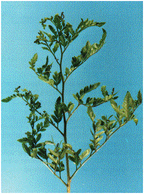
Figure 1A: Necrosis of tips of fern leaflets typical of a light infection of leatherleaf fern anthracnose. [51k]
When the infected frond grows and expands, it appears severely burned or scorched and cannot develop normally. Lesions may appear at or near the base of petioles. The pathogen appears incapable of rhizome infection under natural conditions. Also, mature fern foliage does not appear to be susceptible.
Since the initial symptoms of the disease appear in the understory of the existing mature fern canopy, early stages can be overlooked. When infected fronds unfurl and reveal their damaged condition, the pathogen is already well established in that locale. Symptoms of anthracnose are easily confused with fertilizer or other chemical injury. Infection centers are localized where the disease first appears. These infection centers are usually a few meters in diameter when first noticed, but can enlarge to eventually encompass entire fields.
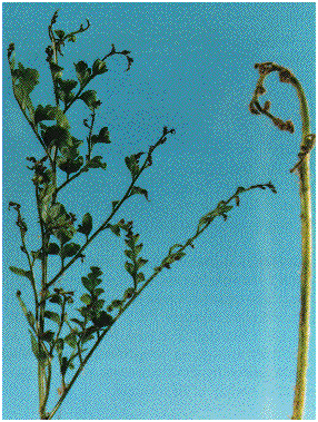
Figure 1B: Severe necrosis of fern fronds in advanced stages of anthracnose. [48k]
DISEASE DEVELOPMENT:
Anthracnose disease development is enhanced by hot, humid, rainy weather. Colletotrichum spores (conidia) enveloped in a slimy matrix are produced within acervuli (fungal fruiting structures). The spores are well-designed for spread by water splash, but can be transported by wind in the form of dry spore masses (Bailey et al. 1992). Inoculum can be easily transported by adhering to hands, tools, clothing, animals, insects, or by moving infected ferns from place to place.
Once conidia come in contact with susceptible young leatherleaf fern tissue, germination will occur in the presence of water. Conidia penetrate host tissue by way of a microscopic structure called an appressorium. Pathogenicity tests conducted by UF-IFAS plant pathologists have shown disease symptoms appearing within 4-5 days after inoculation. These same tests revealed heavy inoculum production on the infected fronds. Based on present knowledge of the disease cycle and of other Colletotrichum diseases, additional inoculum sources are likely in the soil or at the ground line, but early research does not suggest additional inoculum is produced there.
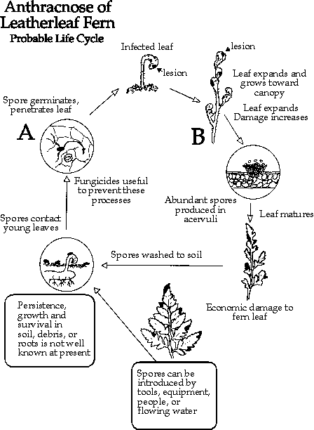
Colletotrichum can survive as conidia, hyphae, appressoria, and possibly sclerotia or hyphal mats (thick-walled, dark masses of vegetative cells) in infected plant debris on or in the soil (Agrios 1988, Bailey et al. 1992). So far, no sexual reproductive stage has been discovered for the causal agent of leatherleaf fern anthracnose. A probable disease cycle of leatherleaf fern anthracnose is presented (Fig. 2).
PREVENTION:
The biology of the leatherleaf fern and its commercial production present a serious challenge to developing an effective control strategy. Prevent movement of this pathogen into uninfected ferneries by limiting access and implementing strict decontamination procedures to delay or prevent infection, especially in isolated ferneries. Schedule activities to avoid the necessity of visiting uninfected areas after visiting infected areas. Activity in an infected area should be scheduled for the end of the work day. Infected areas within a femery should be well marked and avoided by personnel and vehicles except when applying fungicides or other disease management practices.
Decontamination of personnel, equipment, and vehicles is of paramount importance when traveling from one fernery to another. Quaternary ammonium disinfectants are recommended for decontamination. Products such as Galloway GX 1027(1) hand soap-disinfectant used according to label directions can be used for skin decontamination. Quaternary ammonium detergent-disinfectants such as (listed below) mixed with water according to label directions can be used to inactivate anthracnose inoculum on inanimate surfaces such as tools, vehicles, tires, footwear, clothing, and gloves.
- Galloway Gallex 900(1),
[Galloway Chemical Division, 6414 - 125th Ave. N., Largo, FL 34643,
Phone (813) 531-3375, (800) 445-1143],
Green-Shield(2)
[Whitmire Research Labs, 3568 Tree Court, St. Louis, MO 63122,
Phone (800) 325-3668],R.D.-20(3)
[R.D. & Associates, Inc., P.O. Box 1616, Pomona, CA 91769],Prevent(4)
[The Buffalo Co., 6404 Cannel Road, Suite 312, Charlotte, NC 28226], andZepamine A(5)
[ZEP Manufacturing, Atlanta. GA 30301]
Rhizomes should be obtained from anthracnose-free sources when planting new fem beds.
Irrigation should be applied to minimize the dew period. Prolonged wetting encourages inoculum production and enhances infection.
Leatherleaf fern or cut foliage originating from any outside source should be kept well away from ferm production areas. Infected fronds, clippings, cuttings, or debris should be disposed of far from the femery location, or burned, to destroy fungal inoculum and prevent disease spread.
CONTROL:
Early fungicide trials by UF-IFAS researchers have been discouraging (Strandberg 1994, 1995a, 1995b). At rates labeled for foliar spray application, none of the EPA-registered fungicides were especially effective. Fungicidal treatments with EPA-registered products at the shortest intervals pemmitted should be directed at infected fern and plant debris on the ground. Fungicidal spot treatments of newly diseased areas within the femery should include a thoroughly treated 20 foot buffer area around each location. Chemigation (dispensing agricultural chemicals through an irrigation system) has not been tested. More fungicide trials and disease management investigations by IFAS scientists are in progress to determine which compounds and methods provide the most effective control of this disease.
More drastic cultural methods which may reduce an anthracnose
problem are:
- 1) complete mowing and debris removal from infected
areas;
- 2) destruction of all aboveground fern tissues and
accompanying inoculum with flameweeding equipment;
- 3) application of competitive and antagonistic fungi and
bacteria to suppress the anthracnose pathogen;
- 4) combinations of the above treatments with and without conventional fungicides.
These drastic treatments can be expected to reduce or obliterate fern yield for a period of time. The effectiveness of these measures has yet to be thoroughly evaluated. A fern grower should employ any of these measures with caution. A regular monitoring program to detect new outbreaks or follow the progress of treated areas is strongly encouraged.
To determine if you have anthracnose in your fernery, please contact one of the following for advice on sampling and sample submission:
- 1. Ms. Linda Landrum, Volusia County Extension Service, 3100 East New York Avenue, DeLand, FL 32724-6497, telephone 904/822-5778.
- 2. Florida Extension Plant Disease Clinic, P. O. Box 110830, Gainesville, FL 32611-0830, telephone 904/392-1795.
- 3. Florida Department of Agriculture & Consumer Services - Division of Plant Industry, Plant Pathology Section, P. O. Box 147100, Gainesville, FL 32614-7100, telephone 904/372-3505 x 143.
- 4. Mr. Austin Tilton, Putnam County Extension Service, 111 Yelvington Road, Suite 1, East Palatka, FL 32131-8892, telephone 904/329-0318.
LITERATURE CITED
- Agrios, George N. 1988. Plant pathology. Academic Press, Inc., San Diego, California. pp. 379-390.
- Bailey, J.A., R.J. O'Connell, R.J. Pring, and C. Nash. 1992. Infection strategies of Colletotrichum species. pp. 88-120. In J.A. Bailey and MJ. Jeger (eds.). Colletotrichum: biology, pathology, and control. CAB International, Wallingford, Oxford, UK.
- Strandberg, J.O. 1994. Efficacy of selected fungicides for control of anthracnose in leatherleaf fern - a preliminary report. Cut Foliage Grower 9(11/12): 1-4.
- Strandberg, J.O. 1995a. Evaluation of foliar fungicide sprays for control of anthracnose in leatherleaf fern. Cut Foliage Grower 10(3/4): 1-4.
- Strandberg, J.O. 1995b. Efficacy of selected fungicide drenches for control of anthracnose in leatherleaf fern. Cut Foliage Grower 10(7/8): 1-4.
*Authors:
Robert Leahze, Plant pathologist, FDACS, Division of Plant Indurstry, P.O. Box 147100, Gainesville, FL 32614-7100
Tim Schubert, Plant pathologist, FDACS, Division of Plant Indurstry, P.O. Box 147100, Gainesville, FL 32614-7100
Jim Strandberg, Professor, Central Florida REC, 2700 E. Celery Avenue, University of Florida/IFAS, Sanford, FL 32771-9608
Bob Stamps, Professor, University of Florida/IFAS, Central Florida REC, 2807 Binion Road, Apopka, FL 32703-8504
David Norman, Assistant Professor, University of Florida/IFAS, Central Florida REC, 2807 Binion Road, Apopka, FL 32703-8504